Image of an arteriole A venule V and lymph vessel L in the Biology Diagrams
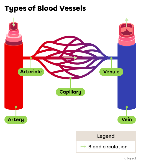
Image of an arteriole A venule V and lymph vessel L in the Biology Diagrams Oxygenated arterial blood circulates through the body via the vascular tree consisting of sequentially smaller arteries, arterioles, and capillary beds. Nutrients and waste exchange between the blood and body tissues occurs at the capillary bed. Venules serve as exit vessels in the capillary bed of various organs and unite to form veins, which return the blood to the heart. The arteriolar wall Blood pumped by the heart flows through a series of vessels known as arteries, arterioles, capillaries, venules, and veins before returning to the heart. Arteries transport blood away from the heart and branch into smaller vessels, forming arterioles. Arterioles distribute blood to capillary beds, the sites of exchange with the body tissues.

Study with Quizlet and memorize flashcards containing terms like Compare and contrast arteries, arterioles, veins, venules and capillaries both functionally and structurally., Explain how blood flows via a pressure gradient., Define systolic and diastolic blood pressure. and more. The different anatomical and physiological features of the arteries, arterioles, veins, venules and capillaries allow each to perform their function correctly. BP and vital perfusion of organs are maintained via a series of mechanisms implicating the baroreceptors, chemoreceptors, RAAS and hypothalamic-pituitary axis.

Blood Vessels: Types, Function & Anatomy Biology Diagrams
The arterioles further subdivide into meta-arterioles. Capillaries . Capillaries are thin-walled vessels composed of a single endothelial layer. Because of the thin walls of the capillary, the exchange of nutrients and metabolites occurs primarily via diffusion. The arteriolar lumen regulates the flow of blood through the capillaries. Venules An arteriole is the smallest division of the arterial network, connecting arteries to capillary beds.. The walls of arterioles contain each of the three layers of blood vessels (tunica intima, media and adventitia), however they are relatively thin and have a narrow lumen. The endothelial cells of arterioles are small with lumen projecting nuclei, the tunica media contains only one to three
The fundamental interorgan variations in microvascular anatomy relate primarily to branching patterns, vessel densities, and the fine structure of capillaries. Below is a brief description of the three structurally and functionally important elements of the microcirculation, i.e., arterioles, capillaries, and venules [14,25-28]. The arterioles further subdivide into meta-arterioles. Capillaries Capillaries are thin-walled vessels composed of a single endothelial layer. Because of the thin walls of the capillary, the exchange of nutrients and metabolites occurs primarily via diffusion. The arteriolar lumen regulates the flow of blood through the capillaries. Venules Venules. Venules (small veins) receive blood from capillaries and lead to veins. venules and arterioles. These smaller vessels connect to 160 arteries and 200 veins. Advertisement. Your most important blood vessel is your body's main artery — your aorta. This is a large artery that carries blood away from your heart and delivers oxygen
